

AM-K70 COLOR DOPPLER ULTRASOUND SCANNER ECHOCARDIOGRAPHY
Introducing the AM-K70 ECHOCARDIOGRAPHY COLOR DOPPLER ULTRASOUND SCANNER, a cutting-edge device revolutionizing diagnostic imaging. With advanced color Doppler technology, this ultrasound scanner delivers precise and detailed images for accurate cardiac evaluations.
Equipped with high-resolution imaging capabilities, the ECHOCARDIOGRAPHY COLOR DOPPLER ULTRASOUND SCANNER AM-K70 provides clear and comprehensive views of cardiac structures and blood flow patterns. Its intuitive interface and user-friendly features streamline the scanning process, ensuring efficient examinations and reliable results.
Designed for versatility, the ECHOCARDIOGRAPHY COLOR DOPPLER ULTRASOUND SCANNER AM-K70 is suitable for a wide range of applications, including echocardiography, vascular imaging, obstetrics, and more. Whether in hospital settings or outpatient clinics, this ultrasound scanner offers unparalleled performance and reliability.
Compact and portable, the AM-K70 is ideal for use in various healthcare environments, providing flexibility and convenience for healthcare professionals. Invest in the AM-K70 ECHOCARDIOGRAPHY COLOR DOPPLER ULTRASOUND SCANNER today and elevate your cardiac imaging capabilities to new heights.
AM-K70 COLOR DOPPLER ULTRASOUND SCANNER ECHOCARDIOGRAPHY
Technical Specifications
| PARAMETER | SPECIFICATION |
| Trolley type full digital color Doppler ultrasonic mainframe | |
| Ultrasonic host operating system | Windows operating system |
| Applications | Abdomen, obstetrics, gynecology, heart, urinary system, small organs, superficial, blood vessels, pediatrics, newborns, musculoskeletal |
| Probes | Convex probe, Tran-vaginal probe, Linear probe, Micro-convex probe, Cardiac probe,4D Volume probe |
| Applications and report | Support abdomen, obstetrics, gynecology, heart, urology, small organs, superficial, blood vessels,pediatrics, etc. advanced measurement software package, report software package, patient management software package |
| carotid artery intima measurement thickness(IMT) | |
| Automatic spectral envelope measurement | |
| Full digital transmission and reception of beam synthesizer | |
| Color Doppler imaging(C) | |
| Pulse Doppler Imaging(PW) | |
| Coherent Contrast imaging(CCI) | |
| Continuous wave Doppler imaging(CW) | |
| B/C/D Real-time three synchronous imaging | |
| Power Doppler imaging(PDI) | |
| Direct power Doppler imaging(DPDI) | |
| M mode imaging | |
| Anatomic M mode imaging | |
| Color Doppler M mode imaging | |
| Elastography | |
| Tissue Doppler imaging(TDI) | |
| Strain rate imaging (SRI) | |
| Tissue harmonic imaging(THI) | |
| Fusion harmonic imaging(FHI) | |
| Speckle Reduce imaging(SRI) | |
| Panoramic imaging | |
| Deflection imaging | |
| Trapezoidal imaging | |
| Adaptive velocity optimization | |
| Free hand 3D | |
| Real time 3D imaging(3D/4D) | |
| DICOM3.0 | |
| Monitor | ≥21.5 inch,high definition ultrasonic display |
| ≥10.4 inch touch screen | |
| Physical clipboard | save the image on the left side of the screen, which can be directly saved ordeleted. |
| The system has the function of on-the-spot upgrade | |
| Presupposition | for different inspection of the viscera, preset the inspection conditions for thebest image, reduce the adjustment of the operation, and the commonly used external adjustment and combination regulation. |
| Probe interface | 4 |
| Chinese and English System, Chinese and English input, optional | |
| Depth | ≥360mm |
| Extended imaging | |
| Probes | |
| Convex probe | Fundamental Frequency:2.0MHz/2.3MHz/2.5MHz/3.0MHz/3.5MHz/4.0MHz/4.6MHz/5.0MHz/5.4MHz,
Harmonic Frequency: 4.0MHz/4.6MHz/5.0MHz |
| Linear probe | Fundamental Frequency:4.0MHz/4.6MHz/5.0MHz/6.0MHz/7.0MHz/8.0MHz/9.2MHz/10.0MHz/12.0MHz/13.3MHz,
Harmonic Frequency: 8.0MHz/9.2MHz/10.0MHz |
| Trans-vaginal probe | Fundamental Frequency:3.0MHz/3.5MHz/4.0MHz/5.0MHz/5.4MHz/6.0MHz/7.0MHz/8.0MHz/10.0MHz
Harmonic Frequency: 6.0MHz/7.0MHz/8.0MHz |
| Micro-convex probe | Fundamental Frequency:3.0MHz/3.5MHz/4.0MHz/5.0MHz/5.4MHz/6.0MHz/7.0MHz/8.0MHz,
Harmonic Frequency: 6.0MHz/7.0MHz/8.0MHz |
| Cardiac probe | Fundamental Frequency:1.7MHz/1.9MHz/2.1MHz/2.5MHz/3.0MHz/3.4MHz/3.8MHz/4.2MHz/5.0MHz,
Harmonic Frequency: 3.4MHz/3.8MHz/4.2MHz |
| 4D Volume probe | Fundamental Frequency:2.0MHz/2.5MHz/3.0MHz/3.3MHz/3.7MHz/4.0MHz/5.0MHz/6.0MHz,
Harmonic Frequency: 4.0MHz/5.0MHz/6.0MHz |
| 2D imaging mode | |
| Gain | 0-100,Step 2 adjustable |
| TGC | 8 segment adjustable |
| Maximum focus point | ≥7, which can be moved throughout the whole process |
| Speckle reduction | 0-5,5 level |
| Space Synthesis | 0-2,2 level(Liner probe: 3 level, cardiac probe:0) |
| Dynamic | 30-180,35 level,step 5 adjustable |
| Line density | low、middle、high,3 level |
| Frame correlation | 0-4,4 level |
| Noise reduction | 0-5,5 level |
| Edge Enhancement | 0-5,5 level |
| Sound power | 2-10, 9 level |
| Grey scale | 0-67, 67 level |
| False color | 0-67,67 level |
| Image style | Soft-Comparison,2 level |
| The screen has real-time display of voice power, probe frequency, dynamic range, pseudo color,gray scale and other 11 parameters can be adjusted | |
| Color Doppler imaging mode | |
| Blood gain | 0-100,Step 2 |
| Parameter display | Velocity、Variance |
| B-Restrain(B/W restrain) | 0-7, 7 level |
| Speed Through | 0-8, 8 level |
| Sampling number | 6-24, 7 level |
| Blood flow preferred | 0-8, 8 level |
| Filtering | 1-6, 6 level |
| Sound power | 2-6, 4 level |
| Noise reduction | 0-4, 4 level |
| Smooth treatment | 0-4, 4 level |
| Frame correlation | 0-6, 6 level |
| Chromatography(Blood flow graph) | 0-37, 37 level |
| Line density | Low-Middle-High, 3 level |
| Frequency | 4 level adjustable |
| Velocity:Minimum 0.4K,Maximum 40.5KConvex probe:0.4K-4.3K-38.5K
Linear probe:0.4K-14.7K-39.0K Trans-vaginal probe:0.4K-7.8K-39.7K Volume probe:0.4K-4.2K-34.8K Micro-convex probe:0.4K-10.3K-40.5K cardiac probe:0.4K-7.8K-39.7K |
|
| PS: The frequency of the probe changes and the frequency value changes | |
| PS: Frame rate changes with speed | |
| Pulse wave Doppler (PW) | |
| Gain | 0-100,Step 2 |
| Spectrum envelope function | real time automatic spectrum envelope, manual spectrum envelope, andother modes. The system automatically analyses and displays various data such as PSV, EDV, RI, PI, S/D, ACC, HR and so on. Can wake up or close |
| Sample volume | 0.5mm~30mm |
| Blood angel | -75—75 degree,Step 5 |
| False color | 0-67, 67 level |
| Dynamic range | 20-40, 4 level |
| Filter | 0-9, 9 level |
| Smooth treatment | 1-4, 4 level |
| Sound power | 2-5, 4 level |
| Volume | 0-100, 10 level,Step 10 |
| Audio filtering | 0-4, 4 level |
| Base line | -1.0~1.0 |
| Grey map | 0-67, 67 level |
| Scan velocity | 100-500, 6 level |
| PRF:Minimum 0.5K,Maximum 87.5KConvex probe:0.5K-4.3K-63.3K
Linear probe:0.5K-14.5K-78.4K Trans-vaginal probe:0.5K-8.1K-78.4K Volume probe:0.5K-4.2K-53.8K Micro-convex probe:0.5K-10.3K-81.1K cardiac probe:0.5K-4.3K-87.5K |
|
| Frequency | 4 level |
| PS: The frequency of the probe changes and the PRF value changes | |
| PS: The frequency of the probe changes and the frequency value changes | |
| Continuous Wave Doppler (CW) | |
| Support probe | Cardiac probe |
| Adjustment of B mode parameters is switchable | |
| Gain | 0-100,Step 2 |
| Sampling line position is adjustable | |
| PRF | 0.9K~36.1K |
| Baseline | -1.0~1.0 |
| Blood angel | -75~75 degree |
| Grey map | 0-67 |
| Scan velocity | 100-300 |
| False color | 0-67 |
| Dynamic range | 20-40 |
| Filtering | 0-9,9 level |
| Smooth treatment | 1-4 |
| Frequency | 2.0MHz/2.3MHz/2.5MHz/3.0MHz,4 level adjustable |
| Sound power | 2-5 |
| Volume | 0-100 |
| Audio Filtering | 0-4 |
| Anatomical M imaging | |
| Support probe | Convex probe, Linear probe, Cardiac probe |
| Adjustment of B mode parameters is switchable | |
| Gain | 0-100,Step 2 |
| M Sampling line angel is adjustable | |
| M Sampling line length is adjustable | |
| Sampling line | 3,Can be displayed or hidden separately |
| Blood flow M model (MC) | |
| Adjustment of B mode parameters is switchable | |
| Gain | 0-100,Step2 |
| MC Sampling line angel is adjustable | |
| MC Sampling line length is adjustable | |
| Frequency | 4 level |
| Sampling number | 6-24 |
| Speed through | 0-8, 8 level |
| Scan velocity | 150-500 |
| Frame correlation | 0-6, 6 level |
| Filtering | 1-6,6 level |
| Blood flow preferred | 0-8,8 level |
| Smooth treatment | 0-4,4 level |
| Map | 0-37, 37 level |
| Elastography | |
| Adjustment of B mode parameters is switchable | |
| Gain | 0-100,Step 2 |
| B/E, Double real-time display on the same screen | |
| Probe displacement curve display | Up/Down |
| Pressure indicator bar display | |
| Frequency | 8-9 level,Adjustable;According to the probe display |
| Noise reduction | 0-2, 2 level |
| Frame correlation | 0-3, 3 level |
| Comparison | 0-13, 13 level |
| False color | 0-3, 3 level |
| Don’t support cardiac probe | |
| Tissue Doppler imaging (TDI) | |
| Support probe | Cardiac probe |
| Adjustment of B mode parameters is switchable | |
| Gain | 0-100,step 2 |
| ROI area adjustable | |
| Sampling number | 6-24 |
| Velocity | 0.4K-8.0K |
| Frame correlation | 0-6,6 level |
| Tissue preferred | 0-7, 7 level |
| Frequency | 2.0MHz/2.3MHz/2.5MHz/3.0MHz |
| Support color reversal | |
| Strain rate imaging | |
| Support probe | Cardiac probe |
| Adjustment of B mode parameters is switchable | |
| ROI area adjustable | |
| Gain | 0-100,Step 2 |
| Sampling number | 6-24,6 level |
| Axial average | 1-4, 4 level |
| Velocity | 0.4K-8K |
| Frame correlation | 0-6, 6 level |
| Tissue optimization | 0-7,7 level |
| Panoramic imaging | |
| Support probe | Linear probe |
| Speckle Reduction | 0-5, 5 level |
| Deflection imaging | |
| Support probe | Linear probe |
| Adjustment of B mode parameters is switchable | |
| Deflection angel | 8 level |
| Speckle reduction | 0-5, 5 level |
| Dynamic rate | 30-180,Step 5 |
| Line density | low-middle-high,3 level |
| Frame Correlation | 0-4, 4 level |
| False color | 0-67, 67 level |
| Image style | Soft-Comparison,2 level |
| Noise reduction | 0-5, 5 level |
| Edge Enhancement | 0-5,5 level |
| Sound power | 2-10,8 level |
| Grey map | 0-67,67 level |
| Trapezoidal imaging | |
| Probe support | linear probe |
| Adjustment of B mode parameters is switchable | |
| Deflection angel | 8 level |
| Speckle reduction | 0-5, 5 level |
| Dynamic rate | 30-180,Step 5 |
| Line density | low-middle-high,3 level |
| Frame Correlation | 0-4,4 level |
| False color | 0-67, 67 level |
| Image style | Soft-Comparison,2 level |
| Noise reduction | 0-5, 5 level |
| Edge Enhancement | 0-5,5 level |
| Sound power | 2-10,8 level |
| Grey map | 0-67, 67 level |
| Space Synthesis | 0-2, 2 level |
| Freehand 3D imaging | |
| Support probe | convex probe, linear probe |
| Display model | 4 pictures |
| Image Rotation X/Y/Z Axis | |
| Multi-slice Visibility | |
| Real-time 4D imaging | |
| Support probe | 4D volume probe |
| Adjustment of B mode parameters is switchable | |
| Gain | 0-100,Step 2 |
| Display model | one image、two images、four images |
| Image Rotation | X/Y/Z Axis |
| Multi-slice Visibility | |
| Light&Shade inversion | |
| Smooth | 0-4, 4 level |
| Threshold level | 0-129, Step 3 |
| Transparency | 1-509,Step 10 |
| Render type | 4 kinds,Surface、maximum、minimum、perspective |
| Extended Imaging | |
| Gain | 0-100,Step 2 |
| TGC | 8 segment adjustable |
| Maximum focus point | ≥7, which can be moved throughout the whole process |
| Speckle reduction | 0-5,5 level |
| Space Compound | 0-2,2 level(Linear probe: 3 level,don’t support cardiac probe) |
| Dynamic range | 30-180,35 level,Step 5 |
| Line density | Low、Middle、High,3 level |
| Frame correlation | 0-4,4 level |
| Noise reduction | 0-5,5 level |
| Edge enhancement | 0-5,5 level |
| Sound power | 2-10, 9 level |
| Grey map | 0-67, 67 level |
| False color | 0-67,67 level |
| Image style | Soft-Comparison,2 level |
| Extended level: Maximum 72 levelConvex probe:9 level
Trans-vaginal probe:72 level Micro-convex probe:29 level Cardiac probe:40 level 4D Volume probe:17 level |
|
| PS:The screen has real-time display of voice power, probe frequency, dynamic range, pseudocolor, gray scale and other 11 parameters can be adjusted | |
| PS: When the probe scan range reaches the maximum, the space synthesized is 0. | |
| Measurement and analysis function | |
| General measurement: Distance, area, ellipse, cross line, angle, distance ratio, volume, Volume(ellipse), area ratio, diameter, joint angle | |
| Cardiac | Automatic spectrum envelope、LV、Main Pulmonary artery diameter、RVEDd、RVEDs、LVM、LAV、HR、MVF、AO、AR、LVOT、TVF、Pulmonic valve、Pulmonary vein、RV、Doppler fetal heart sound、LVET、LVM、LVMI、AV |
| Vascular | carotid intima (IMT), length stenosis ratio, area stenosis ratio, IMT (back wall), IMT (front wall) |
| OB | Fetal routine、AFI、TW、GS、CRl、OFD、HL、ulna、NT、Fibula、Nbonel、Radial、Tibia |
| GYN | uterus、cervix、corpus uteri/cervix uterus、left ovarian vein、right ovarian vein、dominant follicle、intima thickness |
| Urology | prostate、residual urine、left kidney、right kidney、left suprarenal vein、right suprarenal、left testis、right testis、left seminal vesicle、right seminal vesicle |
| Abdomen | liver、CHD、partal vein diameter、cholecyst、CBD、pancreas、spleen、Internal diameter of abdominal aorta、kidney |
| Small parts | Thyroid |
| Software package | Measurement package、Software package、Medical records managementsoftware package |
| Graphic and text management system | |
| Host build in 2 hard disk(SSD 120+1T),Start fast and stable | |
| Movie playback | ≥1200 frames |
| Internal file information management system: can record patient number, name, check number,check date and so on, and can be searched and managed by numbering, checking number, name
and so on. |
|
| Type of report | 16 |
| One key fast report graphic and text management | |
| Interface | |
| USB interface | 4 |
| HDMI interface | 1 |
| RJ-45 interface | 1 |
| Grounding wire interface | 1 |
| DVD RW | 1 |
| Configuration | |
| Trolley type full digital color Doppler ultrasound diagnostic system | |
| Probe | convex array probe (standard), linear probe (optional), Trans-vaginal probe (optional), cardiac probe (optional), 4D volume probe (optional) |
| ≥13 quick adjusting knobs | |
| Technology, after-sales service and other requirements | |
| After acceptance, the warranty is free for two years(Provide manufacturer warranty certificate) | |
| Manufacturer has ISO13485 certification and EU CE certification. | |
Based on 0 reviews
Be the first to review “AM-K70 COLOR DOPPLER ULTRASOUND SCANNER ECHOCARDIOGRAPHY”
Related products
-
MEDICAL
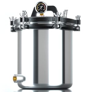
Autoclave 18L
Experience superior sterilization with our Autoclave 18L . Designed for efficiency and reliability, our autoclave ensures thorough sterilization of medical equipment, laboratory instruments, and more. With its spacious 18-liter capacity, this autoclave accommodates a wide range of items, making it ideal for medical facilities, laboratories, and dental offices. Trust in our autoclave to provide consistent, high-quality sterilization results, ensuring the safety and integrity of your instruments. Invest in the peace of mind that comes with superior sterilization – choose our Autoclave 18L for your sterilization needs.
SKU: n/a -
MEDICAL
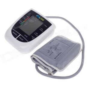
Fully Automatic Blood Pressure Monitor – Jziki
Track your blood pressure with confidence using the Fully Automatic Blood Pressure Monitor by Jziki. With its advanced technology and user-friendly design, this monitor offers accurate readings at the touch of a button. Stay informed about your health journey with ease, thanks to its memory function for tracking progress. Compact and portable, it’s perfect for use at home or on the go. Trust in Jziki for reliable health monitoring. Invest in your well-being today with the Fully Automatic Blood Pressure Monitor by Jziki.
SKU: n/a -
MEDICAL
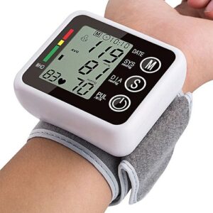
Blood Pressure Monitor -Wrist
Stay on top of your health with our Wrist Blood Pressure Monitor. Compact and convenient, this device offers accurate blood pressure readings with ease. Perfect for home use or on-the-go monitoring, our wrist blood pressure monitor provides reliable results whenever you need them. Take charge of your well-being with our user-friendly device, designed to make tracking your blood pressure effortless. Trust in our Wrist Blood Pressure Monitor for reliable readings and peace of mind.
SKU: n/a -
MEDICAL
Analytical balance
Unlock precision in your laboratory work with our cutting-edge Analytical Balance. Elevate your scientific endeavors with accuracy and reliability. Our analytical balances offer unparalleled precision, ensuring meticulous measurements for your critical experiments. Experience the epitome of balance technology, crafted for accuracy in every detail. Trust in our Analytical Balance to deliver consistent and reliable results, setting the standard for excellence in laboratory instrumentation. Revolutionize your research with the precision you can depend on. Explore the future of analytical precision today!
SKU: n/a

There are no reviews yet.